 |
| Mosquito dissection under a microscope. |
Mosquito dissection - a technique to determine the role of mosquitoes in transmitting malaria (Part 2)
Review Part 1
Dissecting mosquitoes to determine the stages of mosquito egg development The mosquito dissection technique can also determine the developmental stages of mosquitoes based on specific criteria when observing the egg strings. To determine these stages, it is crucial to identify the physiological condition of the collected female mosquitoes. Previously, the physiological condition of mosquitoes was determined using the sella index, ranging from sella I to sella VII. Presently, the physiological condition of mosquitoes is categorized into the following four stages: The mosquito dissection technique serves as a valuable method in determining the developmental stages of mosquitoes, primarily by observing specific criteria evident in the egg strings. Assessing these stages requires accurate identification of the physiological condition of the collected female mosquitoes. Previously, the physiological condition of mosquitoes was assessed using the sella index, which categorized conditions from sella I to sella VII. However, contemporary methods categorize the physiological condition of mosquitoes into four distinct stages: Hungry mosquitoes, Freshly-fed or non-gravid mosquitoes, Semi-gravid mosquitoes, Gravid mosquitoes. This refined classification system aids researchers in accurately discerning and analyzing the developmental stages of mosquito eggs during dissection. The abdomens of females from unfed to gravid are as follows: - 'Unfed': females show collapsed abdomens, with the stomach empty and the ovaries taking up no more than one third of the abdomen. - 'Freshly fed': the abdomen is greatly swollen and red with blood. only the terminal 2-3 ventral segments and 4 dorsal segments are not red, occupied instead by the ovaries. - 'Semi-gravid': the blood meal is greatly reduced and very dark, occupies between the 2-3 ventral segments and 1-2 dorsal segments, closest to the thorax. The ovaries take up most of the abdomen. - 'Gravid': the blood meal is nearly, if not fully digested. The dark colouration is reduced to a narrow, black line on the still swollen abdomen. The ovaries take up most of the abdomen. In some mosquito species, the buoyant floats on either side of the eggs can be observed through the abdominal wall. The female is now ready to oviposit. After determining the physiological state of the mosquito, mosquito dissection is performed. Observing the ovaries under 40x magnification can record the developmental stages of eggs within in the ovarian follicles from immature eggs to mature eggs. Typically, each ovary holds around 75 egg strings or more. Within each egg string, there are two to three egg follicles, each containing a single immature egg. As a result, an average ovary houses approximately 200 eggs. Mosquito eggs progress through five developmental stages. These stages play a crucial role in the life cycle of mosquitoes, ultimately leading to their hatching and further development into larvae. - Stage 1: The egg follicles are round with no visible nuclei. The oocyte is small and indistinguishable from the nurse cell nucleus. This stage characterizes in newly emerged female mosquitoes that have not yet fed on blood. - Stage 2: The egg follicles are oval-shaped and exhibit visible nuclei. The nuclei occupy half of the egg follicle, and the egg nuclei are observable. Stage 2 can be further divided into three periods: pre-period, mid-period, and post-period. Pre-period: Some visible yolk granules surround the egg nucleus but are only observable under a microscope at 40x magnification. This period is typically found in female mosquitoes developing their eggs for the first time. Mid-period: The yolk granules occupy a quarter of the egg follicle and are observable under less than 10x magnification. This period is seen in female mosquitoes that have just finished a blood meal and are developing their initial batch of eggs. Post-period: Numerous visible yolk granules occupy up to half of the egg follicle, and the oocyte nucleus are still observable. This period is common in blood-fed mosquitoes that have laid eggs at least once or in newly developing egg batches. 
Feeding mosquitoes with mice. - Stage 3: The yolk granules occupy about three forths of the egg bulk and made the oocyte nucleus hardly visible. - Stage 4: The yolk granules occupy the entire egg follicle, and the egg follicle elongates, resembling a string of beads. - Stage 5: The eggs have fully developed and have floats on both sides. Dissecting mosquitoes to find the parasitic oocyst in the midgut and sporozoite in the salivary glands Why do sporozoites in the mosquito's salivary glands play a role in disease transmission? The life cycle of the malaria parasite involves two stages: the asexual development stage in the human body and the sexual development stage within the mosquito. When a female Anopheles mosquito bites an infected human, it injects male and female gametocytes into the human's bloodstream. These gametocytes then develop into male and female gametes. A female gamete matures into a female gametocyte, while a male gamete develops into multiple mature male gametocytes through exflagellation. Fertilization occurs when male gametocytes unite with several female gametocytes, forming a zygote. The zygote becomes motile and transforms into a mobile egg called an ookinete. This egg penetrates the mosquito's midgut wall, transforming into an oocyst. As the oocyst matures, it contains around 10,000 sporozoites. When the oocyst ruptures, these sporozoites gather in the mosquito's salivary glands. Upon biting a human, the sporozoites from the mosquito's salivary glands are injected into the human body, continuing the life cycle through the asexual development stage of the malaria parasite in the human body, ultimately causing malaria. Therefore, when dissecting the salivary glands of female Anopheles mosquitoes and discovering the presence of malaria parasite sporozoites, it's possible to determine the mosquito's role in disease transmission at that point. In other words, it serves as the local malaria disease transmitter. Dissecting mosquitoes to identify the oocyst stage of the parasite. The mosquito dissection technique serves the purpose of locating the oocysts of the malaria parasite residing in the mosquito's midgut. Following a female mosquito's blood meal from an infected individual, male and female gametocytes of the malaria parasite combine in the mosquito's midgut, forming oocysts. These oocysts represent the initial stage of the parasite's development within the mosquito. Through mosquito dissection and microscopic examination of their midgut, typically at magnifications ranging from 10 to 40 times, researchers can identify the presence of these malaria parasite oocysts inside the mosquito's body. 
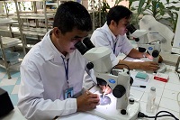
A mosquito dissection typically takes place within a few minutes. In the photo: Mr. Phan Chau Do (left) and Mr. Pham Quang Luan, staff members of
Entomology Department of IMPE-QN, meticulously dissecting mosquitoes. Dissecting mosquitoes to determine the sporozoite stage of the parasite. Besides detecting oocysts, mosquito dissection serves another crucial purpose: identifying sporozoites of the malaria parasite residing in the mosquito's salivary glands. As the oocysts of the malaria parasite mature within the mosquito's midgut over time, they develop into sporozoites. These sporozoites then migrate and concentrate within the mosquito's salivary glands. The dissection method exposes these salivary glands, enabling the direct observation of malaria parasite sporozoites. This technique of dissecting mosquitoes to locate sporozoites plays a pivotal role in determining the disease-transmitting potential of a specific Anopheles mosquito species as a local malaria vector. In this method, upon coating mosquito salivary gland samples with enamel/clear resin, gentle tapping with a dissecting needle uniformly ruptures the salivary gland, releasing sporozoites. Microscopic examination at 40x magnification reveals spindle-shaped sporozoites that refract light, occasionally displaying slow wriggling movements within the slide. Identifying sporozoites or oocysts of the malaria parasite through mosquito dissection necessitates the use of freshly collected mosquito samples. Despite successfully locating sporozoites and oocysts, determining the specific species of the malaria parasite causing the infection may prove challenging. The method of mosquito dissection aimed at finding sporozoites and determining the disease-transmitting role of mosquitoes is both time-consuming and labor-intensive. Moreover, the accuracy and outcomes often hinge on the expertise and skills of entomologists, colloquially known as 'mosquito surgeons'. How is mosquito dissection performed? Dissecting a mosquito is a meticulous surgical procedure owing to the small size of adult mosquitoes. It necessitates specialized equipment, including a binocular microscope and a mosquito dissection table, to magnify the area being examined. Ideally, the dissection and classification of captured mosquitoes should occur on the same day, with morning-captured mosquitoes dissected in the afternoon and night-captured ones dissected the following morning. Before dissection, mosquitoes should be anesthetized using ether or chloroform. The dissection itself takes place on a glass slide under a microscope, utilizing a specified dissection technique. During this process, observations can be made regarding various aspects such as egg maturity, physiological age, egg-laying behavior, and the detection of oocysts in the mosquito midgut, along with sporozoites in the salivary glands. To detect fresh sporozoites, adjust the condenser's light intensity for optimal brightness. Observing shapes resembling aligned, white-colored cut nails displaying a wavelike motion within the salivary glands indicates the presence of sporozoites. However, care must be taken not to mistake these movements for flagellates. Sporozoites are typically 1.5 times longer than flagellates and possess two tapered ends. Suspected sporozoites can be further examined by staining the specimen with Giemsa and using oil immersion microscopy. Under staining, sporozoites typically exhibit a spearhead shape with a red-stained center and violet hue at the edges. Proper staining reveals a red-stained center, blue-stained ends, and a pinkish-violet hue near the center. The identification of sporozoites in the salivary glands serves as a vital determinant of the disease transmission potential of the Anopheles mosquito species in a given locality. The unsung heroes known as 'mosquito surgeons' encompass entomologists and technicians whose silent yet invaluable efforts remain largely unnoticed in the ongoing battle against malaria. While they may not enjoy the spotlight akin to surgical doctors in traditional healthcare systems, their contributions are profound, especially in preventive medicine, notably within the realm of malaria prevention. These dedicated individuals play a pivotal role in identifying mosquito-related issues, specifically in determining the disease transmission roles of Anopheles mosquitoes within local communities. This information forms the bedrock for implementing effective and accurate intervention measures to combat malaria. Admirably, these specialists in the field of 'mosquito surgery' diligently pursue their scientific endeavors, working tirelessly, one mosquito at a time, contributing significantly to the noble cause of scientific advancement. 
|

 Forum
Forum Sitemap
 Contact


 Petechial fever
